
New underwater microscope shows how climate change affects coral reefs
Just last month, Forbes reported Australia’s Great Barrier Reef is struggling to survive. Their article stated, “The Great Barrier reef is a victim of global climate change and its decline is a direct result of a warming planet.” They described how back-to-back bleaching events and a hurricane have left the reef vulnerable. Much of the reef is in danger of dying off.
Popular Science echoed the sentiment on coral bleaching, asserting, “Warning: coral reefs might be a lot more sensitive to global warming than originally predicted.” This article shared research on a coral reef in the South China Sea. Forty percent of this reef died after a bleaching event caused by moderate ocean warming.
The importance of coral reefs
There are many good reasons environmentalists are focused on coral reef preservation. Notably, coral reefs are critical to our planet’s biodiversity. The Coral Reef Alliance states, “Coral reefs are believed by many to have the highest biodiversity of any ecosystem on the planet—even more than a tropical rainforest. Occupying less than one percent of the ocean floor, coral reefs are home to more than twenty-five percent of marine life.”
Coral reefs also protect the shorelines from wave erosion in many coastal areas, create jobs based on tourism, and even provide a source for new medicines. They also act as a hatchery for spawning fish and provide food resources that feed tens of millions of people. According to Scientific American, coral reefs ecosystems contribute an estimated $172 billion a year to the world economy,
Underwater microscope is bringing the lab to the ocean floor
Coral bleaching occurs when corals expel symbiotic algae. It is called bleaching since it is the algae that give corals their color. The bleached corals are still alive, but since the symbiotic algae provide the corals with 90% of their energy, they begin to starve. In its weakened condition, the corals are susceptible to invading algae that smother the coral polyps.
While corals can recover from a bleaching event, back-to-back events often prove fatal. Scientists have documented how the stress of increased temperature connects to the bleaching events, but they have yet been unable to study the bleaching process as it occurs in the ocean at the individual coral polyp level. Typically, the coral is removed from the sea floor, brought to a lab, and is sliced for study under a microscope.
To increase our understanding of corals’ interactions in their natural environment, researchers from Scripps Institution of Oceanography at the University of San Diego have designed an underwater microscope that captures images and videos at the millimeter-level scale. It is the first microscope that can capture corals’ processes as they occur in nature. The New York Times reported, “They developed the tool to study what happens during coral bleaching and its aftermath.”
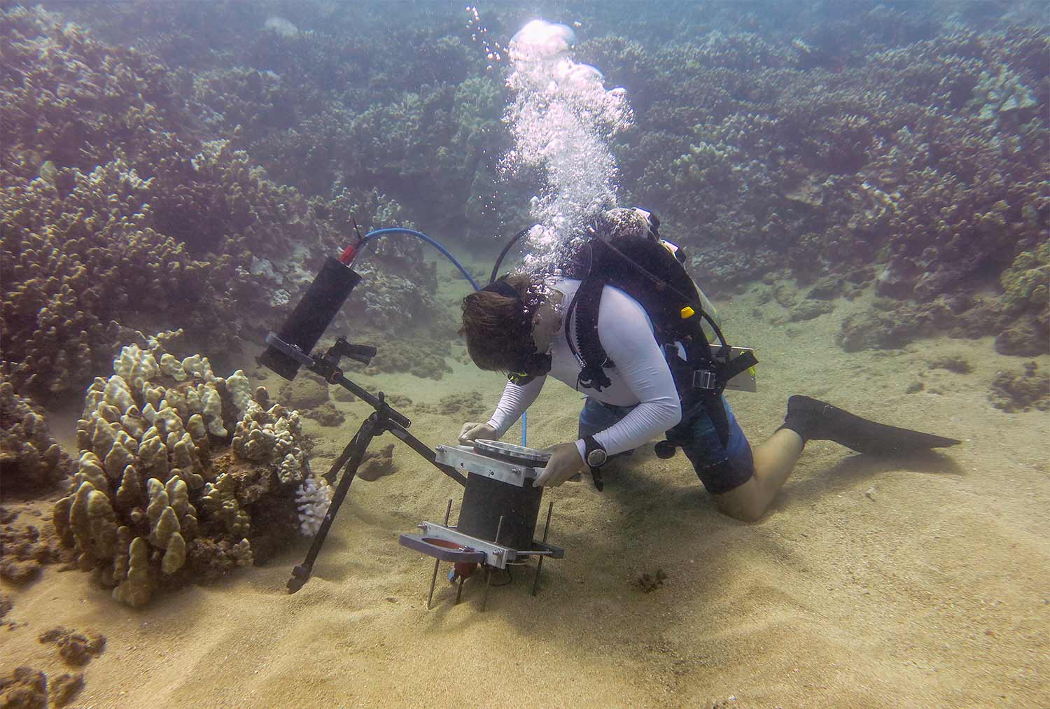
The Benthic Underwater Microscope is positioned to study coral competition. Credit: Jaffe Laboratory for Underwater Imaging/Scripps Institution of Oceanography, UC San Diego
The Benthic Underwater Microscope (BUM) enables divers to capture photos of corals without removing samples to be studied in a lab. Its design is detailed in a paper in Nature Communications titled “Underwater microscopy for in situ studies of benthic ecosystems.” Scientists originally developed the camera system to film plankton but adapted it to monitor coral reef bleaching and algal overgrowth.
“This instrument is part of a new trend in ocean research to bring the lab to the ocean, rather than the ocean to the lab,” added Dr. Tali Treibitz, University of Haifa’s Charney School of Marine Science and co-author of the paper.
The BUM is a two-part system. It combines a microscopic imaging system with an underwater resolution of 2.2 μm and an underwater computer with a diver interface. The imaging system uses a tunable lens consisting of a flexible polymer membrane encasing an optical fluid. The system exerts variable pressure on the encased fluid to change the lens curvature and focal length. The system also has a custom-designed ring of six LEDs for illumination.

The BUM is positioned to study coral competition. Credit: Jaffe Laboratory for Underwater Imaging/Scripps Institution of Oceanography, UC San Diego
“To understand the evolution of the dynamic processes taking place in the ocean, we need to observe them at the appropriate scale,” said Jules Jaffe, the senior author of the study and head of the Jaffe Laboratory for Underwater Imaging at Scripps.
The lens can also scan the optical system’s focal plane through a volume to bring various parts of a subject in focus. Frames can then be combined using image-processing techniques to produce a single image with all parts of the subject in focus. This is commonly known as focus stacking. The image processing for this research paper was performed using MATLAB.
“This underwater microscope is the first instrument to image the seafloor at such small scales,” said Scripps Ph.D. student Andrew Mullen, co-lead author of the study. “The system is capable of seeing features as small as single cells underwater.”
Team observed how bleached coral is overrun by harmful algae
The team discovered that after a bleaching event, dangerous algae invade the coral in a very specific way. The system collected image data by observing bleached corals that were being colonized by invading algae in Maui. As the team captured the algae’s micro-scale spatial patterns, the images were analyzed using statistical analysis. All statistical analyses for the research paper were performed using MATLAB.
“Using the microscope, the research team observed a previously unreported honeycomb pattern of initial algal colonization and growth in areas between the individual coral polyps during coral bleaching. These findings provide insight into a process that Scripps marine ecologist Jennifer Smith, a co-author of the study, called the “succession of algae” where small filamentous algae initially settle on the ridges between coral polyps and eventually smother the living tissue. The images showed that algae are able to actively overgrow living corals during a bleaching event,” stated Phys.org.


コメント
コメントを残すには、ここ をクリックして MathWorks アカウントにサインインするか新しい MathWorks アカウントを作成します。